
Answer in brief
White coral is not automatically dead: bleached coral can retain living tissue, while mortality means tissue loss. Recovery is possible, not guaranteed. More colour in a later photo can contribute to an assessment of the same colony, but does not prove that an entire reef has recovered.
Is white coral necessarily dead?
White coral is not automatically dead, and a bleached colony may recover, although survival is not guaranteed. The key distinction is living tissue: bleaching can leave it in place, while death involves its loss. NOAA’s explanation separates these states. From a distant photograph they may look similar, so look for documented observations of the colony before treating its colour as a verdict on survival.
For a reader comparing images, the useful first question is therefore whether the description concerns colour or tissue. “The colony looks pale” is an observation. “The colony has died” is a biological conclusion that needs supporting evidence. If a caption jumps directly from the first to the second, look for field observations or a monitoring report that explains how the conclusion was reached.
This does not make bleaching harmless. It changes the kind of claim an image can support. A dramatic photograph can show visible stress without telling us the eventual outcome. Treat the picture as a record of appearance at a particular moment, then use its location, acquisition date and accompanying assessment to understand what happened next.
How do living, bleached and dead coral differ?
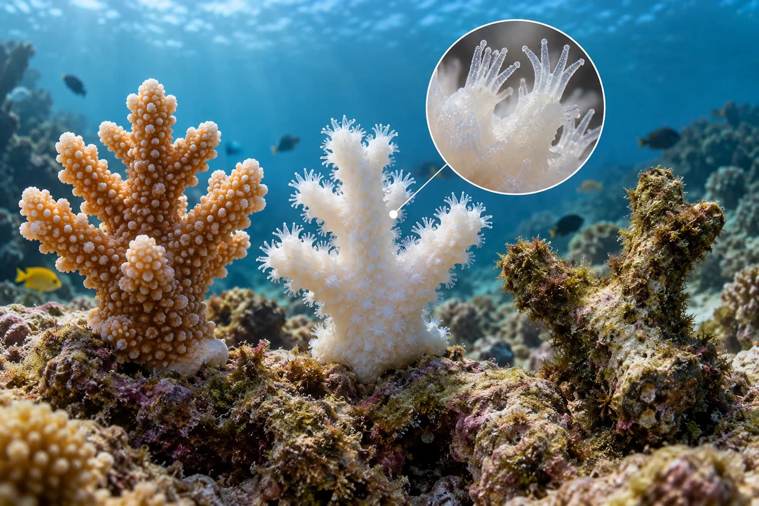
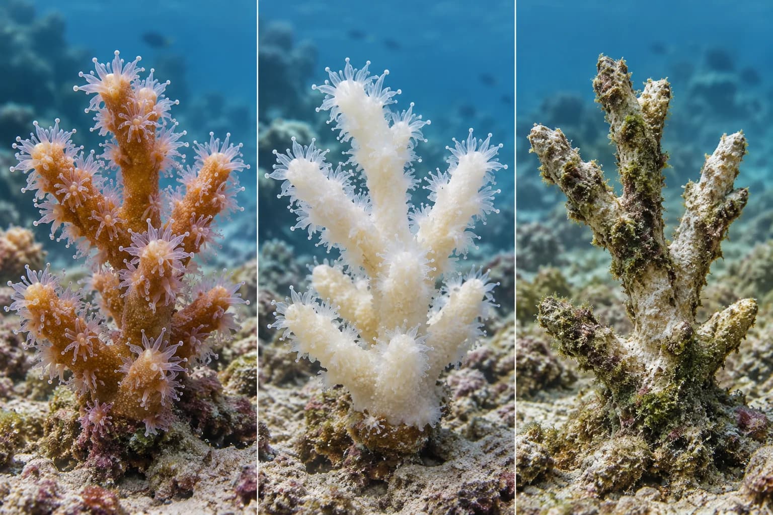
A useful teaching comparison separates pigmented living tissue, bleached living tissue and an exposed skeleton of dead coral. The International Coral Reef Initiative explains that the white skeleton can become visible through bleached tissue and that responses can vary even within a colony. Our cover uses three generic fragments and a tissue inset to make the distinction legible. It does not show stages observed on one individual.
Read the fragments as categories rather than a fixed sequence. A bleached colony may survive, deteriorate or contain areas in different conditions. Putting the fragments from left to right makes an illustration easier to follow; it does not establish that every coral passes through the same outcome. Nor does the apparent size of each fragment represent the share of a reef in that state.
The bare skeleton covered with algae illustrates a possible appearance after tissue loss, not a visual rule for every underwater scene. In an actual report, the observer should identify what is coral tissue and what is growing on an exposed surface. A small inset can explain the distinction, but only observations of the real colony can establish its condition. Never substitute an illustrated polyp for evidence that a photographed coral is alive.
Does the before-and-after pair show the same colony?
A recovery comparison becomes more useful when it follows the same colony or the same defined survey area. Two photographs from different parts of a reef may both be accurate, yet their contrast does not show one place recovering. Start by checking whether the subject can be identified in both images through a documented location, a monitoring identifier or an explicitly described survey method.
Then compare the dates and framing. A wide reef scene and a close view of one branch answer different questions. A later photograph chosen because it contains a colourful surviving colony cannot explain what happened to the colonies missing from the frame. Ask whether the later view covers the original subject and whether the accompanying record accounts for visible losses as well as survivors.
For a personal photo comparison, record the date, the general site and how the subject was matched, without claiming a scientific survey. Keep the originals and describe changes in lighting or camera setup. If the match is uncertain, label the pair as two views rather than a recovery sequence. This preserves the photographs’ value while limiting the conclusion to what their provenance actually supports.
Could the colour change come from the photograph?
Underwater photographs record both the subject and the way it was illuminated and processed. A change in white balance, exposure, shooting distance or use of artificial light can change the apparent colour of the same scene. That is a reason to inspect the image-making conditions before reading a stronger colour as a biological improvement. It is not a claim that every difference is a camera effect.
Look for practical information: were the pictures taken under similar conditions, was a flash used, and were either of the files substantially edited? A comparison can still be informative when these conditions differ, provided the caption acknowledges the difference and the biological assessment has other support. The more tightly the claim depends on colour, the more relevant those details become.
Do not try to repair an uncertain comparison by turning up saturation until the panels resemble an expected story. Preserve the original image and keep any explanatory adjustment separate. An AI illustration is further removed from field evidence: it is designed to express a distinction, not record an observation. Our generated fragments have no location, sampling date or measured recovery period, and their visual clarity cannot supply those missing facts.
Has one colony recovered or the whole reef?
“Recovery” can refer to a surviving colony regaining pigmentation, to living coral cover in a surveyed area, or to broader reef conditions. These are different questions. A photograph of one colourful colony may support discussion of that individual when paired with appropriate observations. It does not, by itself, establish what happened to the whole reef or whether other functions have returned.
Before sharing an optimistic before-and-after image, ask the author to specify the subject and the indicator. Is the comparison about the same colony’s tissue, the extent of living coral in an area, or a longer monitoring record? A clearly defined indicator makes a claim easier to assess. It also allows improvement in one respect to coexist with unresolved damage elsewhere.
The dates in global announcements require the same care. NOAA’s assessment published on June 2, 2026 said the fourth global bleaching event likely concluded in mid-2025. That statement concerns the event’s global classification; it does not certify recovery at every affected site. This article’s October 8, 2026 editorial date does not turn that earlier assessment into a new event. For a particular destination or reef, seek a dated local assessment.
Which evidence answers your recovery question?
Use the comparison table to decide what evidence is missing, rather than to assign a health score to a photograph. Each row separates a visible observation from a broader conclusion. A careful caption may leave a question open. That is more informative than attaching an unsupported final outcome to a picture simply because a side-by-side layout invites a before-and-after story.
If you are assessing a travel operator’s reef images, ask when and where each was taken and whether “recovered” refers to one colony or a monitored area. If you are reading a scientific update, look for the methods and the unit being compared. If you are explaining the biology to a child, the conceptual fragments are useful precisely because their simplified tissue differences can be discussed without claiming a real survey.
Keep three records together when possible: the original image, its descriptive caption and the underlying observation or report. Removing one can change the meaning of the others. A photograph of a survivor placed beside a regional damage headline is not necessarily evidence of regional recovery. The table helps locate that mismatch without pretending that a reader can perform a field diagnosis from a screen.
| What you see or read | What it can suggest | What it cannot establish alone | Useful supporting evidence |
|---|---|---|---|
| A pale or white colony | Possible bleaching | That the coral is dead | Assessment of living tissue |
| Visible tissue on a pale fragment | A distinction from bare skeleton | Its eventual survival | Repeat observations of the same subject |
| Bare skeleton with algal growth | A surface no longer covered by coral tissue | A whole reef’s mortality | Documented tissue assessment and survey scope |
| More colour in a later image | Possible repigmentation | Restored reef function | Matched images and biological observations |
| A colourful reef photograph | The appearance within that frame | Recovery across the site | A defined survey area and monitoring record |
| A global event described as ended | A change in event classification | Recovery of every affected reef | Dated local monitoring |
How to caption the result without overstating recovery
For a genuine paired record, a good caption identifies the subject, dates and observed change, then states the relevant limit. “The same documented colony appears more pigmented in the later image; the accompanying survey reports living tissue” is more precise than “the reef is healthy again.” The first claim can be checked against a record. The second needs a much broader assessment.
For our cover, the correct language is different: an AI-generated conceptual illustration comparing three tissue states. The inset clarifies the intended distinction, while the algal-covered skeleton represents the illustrated dead fragment. None of the fragments documents a diver’s observation, a named reef or a successful restoration project. They should not be described as a real before-and-after photograph.
When evidence is incomplete, retain the uncertainty in the final wording. Say that the coral appears pale, that tissue survival has not been established, or that a site-wide recovery assessment is unavailable. These statements give readers a usable boundary. The most helpful comparison is one that shows what changed, explains what was actually observed and makes clear which conclusion still requires monitoring.
Questions and answers
Is white coral dead, or can it still be alive?
White coral can still have living tissue. Bleaching and tissue death are different states, and survival after bleaching is possible but not assured. Check the accompanying observations rather than treating whiteness as proof of death. The teaching illustration shows categories; it cannot diagnose a real colony or promise its eventual recovery.
Does the return of colour mean the reef has recovered?
It may contribute to an assessment of a particular colony when the subject is matched and biological observations support it. It does not establish recovery across a reef. Ask which indicator, area and dates the claim refers to, and check that colour differences are not being interpreted without considering photographic conditions.
Did NOAA say all coral reefs recovered in 2026?
No. Its June 2, 2026 assessment said the fourth global bleaching event likely concluded in mid-2025. The event’s classification and recovery at an individual site are separate questions. A dated local monitoring report is needed to assess the particular reef you are reading about or planning to visit.
Are the cover’s coral fragments real before-and-after photos?
No. The cover is an AI-generated conceptual illustration showing pigmented living tissue, bleached living tissue and an algal-covered skeleton of dead coral, with an educational tissue inset. It records no named reef or individual colony. The fragments are comparison categories, not an observed recovery timeline or evidence of a restoration result.